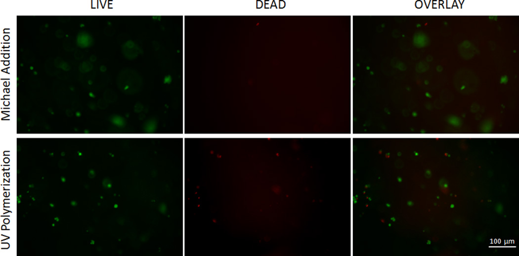
Figure 7.

LIVE/DEAD assay for hydrogels synthesized by Michael Addition and UV polymerization. In the 10× images green fluorescence represents living cells stained by calcein and red fluorescence represents dead cells stained by EthD-1 homodimer. An average live cell percent of 80.9% was observed in hydrogels synthesized using Michael addition, and 52.1% for hydrogels synthesized using UV polymerization.
