Figure 7.

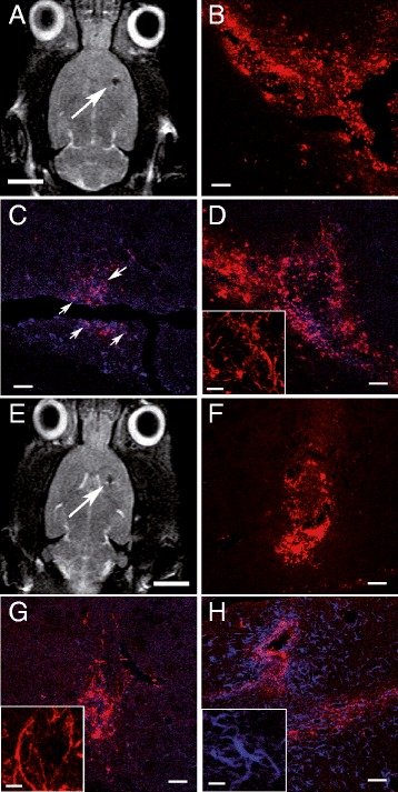
MR imaging of hVM-MNP labeled cells one week and two months after transplantation into hemiparkinsonian rat brains. hVM-MNP-Cy3 labeled cells (3x10⁵ cells) were detected by MRI one week (arrow in A) and two months (arrow in E) after transplantation into the right striatum of 6-OHDA-lesioned rat brains. Analyses by fluorescence microscopy showed that MNPs-Cy3 were clearly visible in red in coronal sections from striatum one week (B) and 2 months (F) after cell transplantation. Transplants were stained for hNu (blue in C, D, G) to detect hVM cells; Ki67 (purple in C, arrows point to Ki67 stained cells, showing a purple color, slightly different from MNPs-Cy3 marked in red); human Nestin (red in D and G, inserts in D and G) and human GFAP (blue in H and insert in H). Scale bars: 5 mm in A and E, 50 μm in B-D, F-H; 20 μm in inserts in D, G-H.
