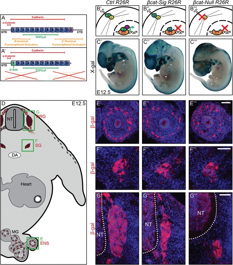
Figure 1.

Total loss of β-catenin leads to a more severe phenotype in the dorsal root ganglia than inhibition of the TCF/Lef transcriptional output of β-catenin. (A) The β-catenin protein consists of 12 Armadillo repeats (numbered boxes), a conserved helix-C (C), an amino-terminal domain (NTD), and a carboxy-terminal domain (CTD) [15]. Colored bars show binding sites for β-catenin interaction partners: red, components of adherens junctions; green, TCF/Lef transcription factors providing DNA binding; orange, transcriptional co-activators. (A’) Diagram presenting β-catenin-dm. D164A and truncation of CTD inhibits association with multiple players of the transcription machinery. However, binding sites for TCF/Lef and components of adherens junctions are preserved. (B’-B”’) Schematic of the functional properties provided by the distinct β-catenin alleles. (B’) β-catenin (green) of control animal induces transcription (green arrow) by binding with TCF/Lef (orange) in the nucleus and recruiting co-transcription factors (purple). Furthermore, it links transmembrane cadherins via α-catenin (yellow) to the actin cytoskeleton (dotted line). (B”) β-catenin-dm (blue) inhibits TCF/Lef-mediated transcription, but preserves cadherin-mediated adhesion. (B”’) Cells of βcat-Null animals lose both TCF/Lef-mediated transcription and cadherin-mediated adhesion. (C’-C”’) In vivo fate mapping of Wnt1-Cre embryos carrying the R26R reporter allele at E12.5. (D) Illustration of a transverse section of an E12.5 control animal displaying in red the neural derivatives of neural crest cells: dorsal root ganglia (DRG); sympathetic ganglia (SG); enteric nervous system (ENS). Green boxes display caption area for subfigures E-G. (E’-G”’) Immunohistochemistry for β-gal on transverse sections. Normal development of the ENS (E’-E”’) and the SG (F’-F”’) can be witnessed in both mutants at E12.5. (G’-G”’). The size of the DRG of βcat-Sig R26R embryos is strongly reduced (G”), whereas DRG of βcat-Null R26R animals are virtually non-existent (G”’). NT, neural tube; DA, dorsal aorta; MG, mid gut. Scale bars: 50 μm. E, embryonic day.
