Figure 2.
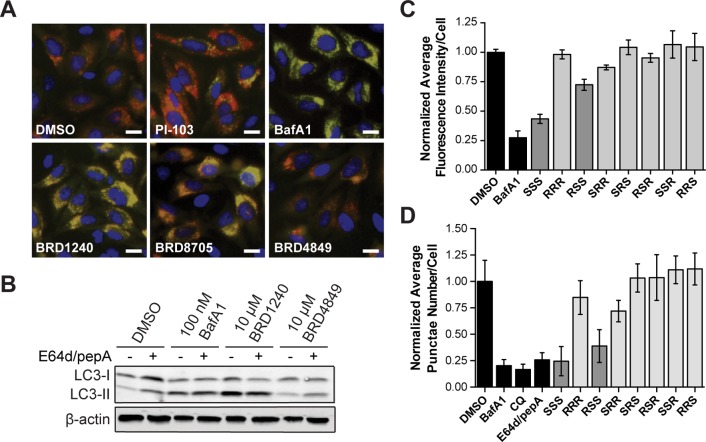
BRD1240 blocks the later stages of autophagy. (A) Representative images from the mCherry-eGFP-LC3 assay following treatment with DMSO, PI-103 (5 μM), BafA1 (100 nM), BRD1240 (10 μM), BRD8705 (10 μM), and BRD4849 (10 μM). Blue (Hoechst 33342), red (mCherry), green (eGFP). Scalar bars represent 10 μm. (B) Western blot for LC3-I to LC3-II shift in HeLa cells treated with BafA1, BRD1240, and BRD4849 ± 10 μg/mL E64d/pepA. (C) Normalized average fluorescence intensity of punctae per cell for BafA1 (100 nM), BRD1240 (SSS) (10 μM), and all seven stereoisomers (10 μM) in HeLa cells in the LysoTracker displacement assay. (D) Normalized average punctae number per cell for BafA1 (200 nM), chloroquine (CQ) (50 μM), E64d/pepA (10 μg/mL), BRD1240 (20 μM), and all seven stereoisomers (20 μM) in HeLa cells in the DQ-BSA assay. In parts C and D data are presented as the average ± SD of three independent experiments, each run in duplicate.

