Figure 5.

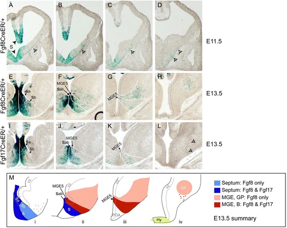
Origins of Fgf lineage cells in the basal ganglia. Xgal stained coronal sections through caudal telencephalons of (A-D) E11.5 Fgf8 CreER/+; ROSA26R, (E-H) E13.5 Fgf8 CreER/+; ROSA26R, and (I-L) E13.5 Fgf17 CreER/+; ROSA26R embryos (Tm E8.5). (M) Diagram illustrating similarities and differences between E13.5 fate maps. In panels (F), (J), and (Mii), Sab indicates that we are unable to distinguish Sa and Sb; the dark blue septal field extending from Sab in (Mii) likely contains cells from both progenitor domains. As shown in (H) and (L), the hypothalamus contains Fgf8 + but not Fgf17 + lineage cells at E13.5. Arrows in (A-D) indicate cells concentrated near the pial surface that appear to be migrating dorsally and caudally from the septum and vMGE. Arrowhead in (L) indicates cells from this pial migratory stream that appear to migrate radially toward the nucleus basalis of Meynert. Lateral striatum cells in the Fgf8 + lineage, indicated in pink in (Mi), are likely be derived from Sb. Abbreviations: S, septum; POA, preoptic area; Hy, hypothalamus; B, nucleus basalis of Meynert. See also Additional file 5: Figure S5.
