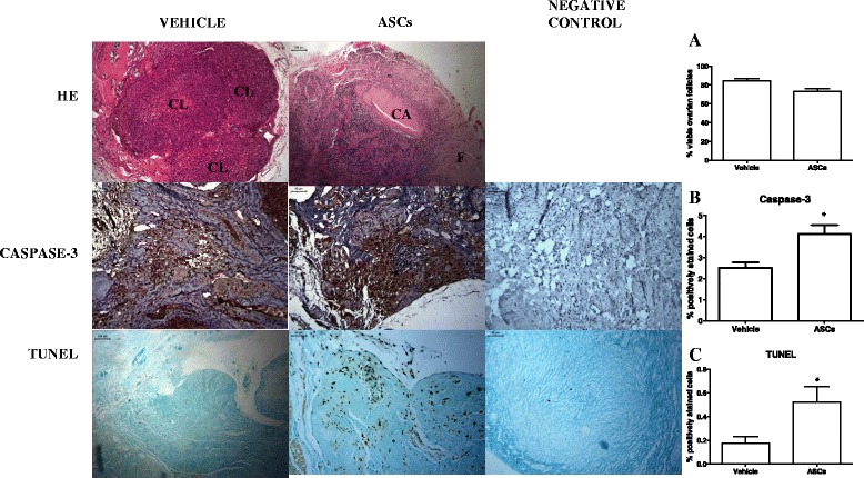
Figure 1.

Photomicrography of cryopreserved ovarian grafts treated with vehicle or ASC 30 days after an autologous avascular transplant. The morphology of the ASCs-treated grafts shows increased fibrosis and more corpora albicans, but the follicle viability was maintained, as evidenced by trypan blue (A) (P >0.05). Immunohistochemistry for apoptosis was performed on the ovarian stroma, and dark brown-stained cells are considered positive. Results are expressed as a percentage of the positive area (arbitrary unity/mm2). Apoptosis increased in both analyses (cleaved-caspase-3 and TUNEL) of the ASC-treated grafts (B and C) (P <0.05). H & E 100x. Immunohistochemistry analysis 200x. ASCs, adipose tissue-derived stem cells; CL, corpora lutea; CA, corpora albicans; F, fibrosis. * P <0.05, unpaired t-test.
