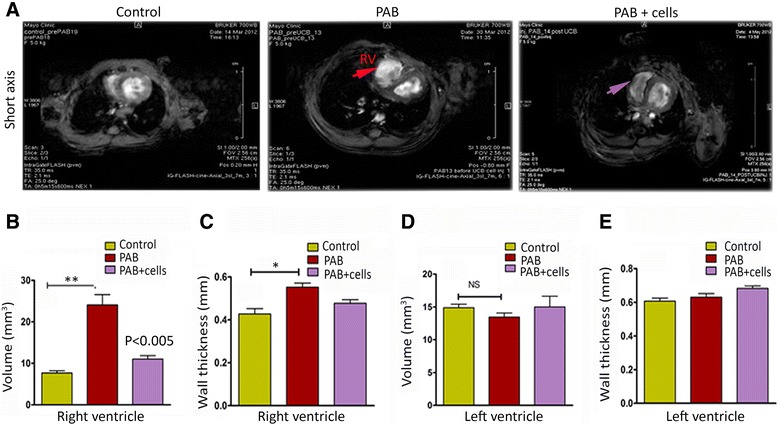
Figure 5.

Myocardial delivery of UCB-MNCs improves RV function and favorable RV remodeling after pulmonary artery banding. (A) Short-axis image obtained from magnetic resonance imaging in the three experimental groups. Pulmonary artery banding (n = 6) produced severe RV dilation and ventricular dysfunction when compared with control (sham) animals (n = 6). Four weeks after cell transplantation, there was a less-pronounced RV dilation in the UCB-MNCs transplanted group (n = 6). (B) Magnetic resonance imaging-derived right ventricular volumes in control, PAB and cell transplanted group. The PAB only group demonstrated an increase in RV volume (** P-value <0.005 versus control). Intramyocardial delivery of UCB-MNCs indicated a reduction in RV volume (P-value <0.005 versus PAB). (C) Right ventricular wall thickness was compared among groups; the PAB-only animals showed a significant increase in RV wall thickness (* P-value <0.05); however, the cell transplanted group demonstrated a smaller reduction of the RV wall thickness. (D, E) No significant changes in LV functions were noticed between the experimental groups. LV, left ventricle; PAB, pulmonary artery banding; RV, right ventricle; UCB-MNCs, umbilical cord blood-mononuclear cells.
