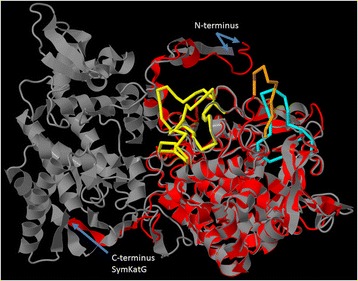
Figure 6.

Superimposition of bacterial and Symbiodinium KatG. Shown are the predicted mature SymKatG1 monomers from Symbiodinium B1 (Mf1.05b, C-score = 0.09, red) superimposed with the crystal structure monomer from Haloarcula marismortui (RCS PDB ID 1ITK_A, grey). SymKatG inserts 1 (yellow), 2 (orange) and 3 (cyan) have been highlighted in the Symbiodinium protein (cf. Figure 5, Additional file 8).
