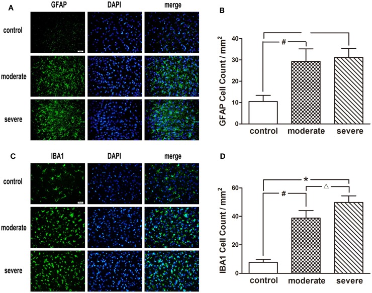
Figure 6.
Immunofluorescence examination in the peripheral area of injured cortex at 72 h after blast-induced traumatic brain injury (bTBI). (A) Astrocytes were stained by GFAP, the nuclei were stained by DAPI (scale bar = 20 μm). (B) Microglias were stained by IBA1, the nuclei were stained by DAPI (scale bar = 20 μm). (C) GFAP positive cell count in the peripheral area of injured cortex at 72 h after bTBI. (D) IBA1 positive cell count in the peripheral area of injured cortex at 72 h after bTBI.

