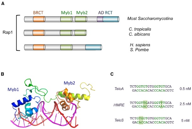
FIGURE 2.

The domain organization of Rap1 and the structure of Rap1DBD-DNA complex. (A) The domain structures of Rap1 from various Saccharomycotina and other species are illustrated. The BRCT, Myb, AD (activation domain), and RCT (Rap1 C-terminal) domains are displayed in different colors. (B) The crystal structure of the Myb1 and Myb2 domains of S. cerevisiae Rap1 (shown in color spectrum from blue to orange) bound to its target DNA (shown in magenta and red; PDB ID: 1IGN). (C) The sequences of the three duplex oligonucleotides bound by ScRap1DBD in a crystallographic study are displayed. The half sites in each oligo are shown in green, and nucleotides that deviate from a canonical half site (5′-GGTGT-3′/5′-ACACC-3′) are shown with a shaded background. The affinities of Rap1DBD for each sequence are shown on the right. Other variant targets (e.g., the site upstream of ribosomal protein genes: AAATGTATGGGTGT) have been reported to have comparable affinities (Idrissi et al., 1998; Idrissi and Pina, 1999).
