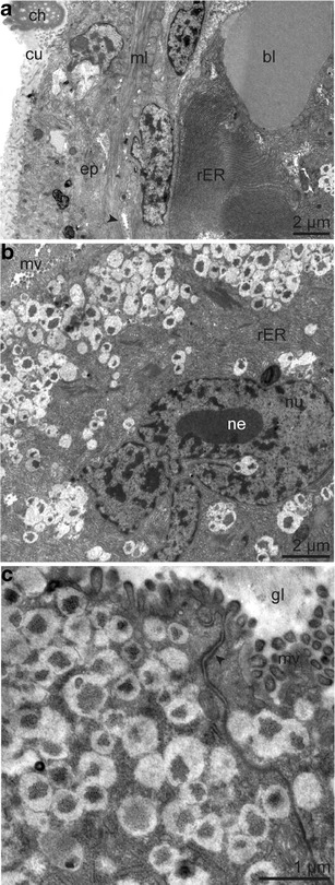
Fig. 10.

Ultrastructure of epidermal multicellular glands of the opisthosoma. a Overview of epidermis, muscle layer with glandular duct (arrowhead) and glandular cell with rER in concentric circles, next to blood lacuna. b Glandular cell with large-lobed nucleus and nucleolus. c Detail of glandular epithelium showing apical junctional complex (arrowhead) and cytoplasm full of electron-light granules containing electron-dark patches. Abbreviations: bl = blood lacuna; ch = chaetae; cu = cuticle; gl = glandular lumen; ml = body wall muscle layer; mv = microvilli; ne = nucleolus; nu = nucleus; rER = rough endoplasmic reticulum
