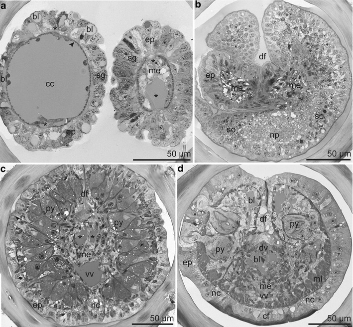
Fig. 4.

Semithin section series of tentacles and forepart. a Left tentacle at distal position with vascularized epidermis overlaying a single-layered myoepithelium (arrowhead) surrounding a central coelomic cavity. Right tentacle at proximal position with mesenchyme filling the coelomic cavity. Each tentacle with two blood vessels (asterisk). b Base of cephalic lobe and of tentacles and beginning of the dorsal furrow; cephalic lobe with the brain consisting of central neuropil and peripheral somata; tentacles with mesodermal strands. c Forepart anterior to the frenulum with densely packed pyriform glands, single ventral nerve cord, and paired dorsal blood vessels (asterisk). d Forepart posterior to the frenulum with pyriform glands loosely distributed from dorsal to lateral and ventral nerve encasing the ciliated field. Abbreviations: bl = blood lacuna; cc = coelomic cavity; cf = ciliated field; ep = epidermis; df = dorsal furrow; dv = dorsal blood vessel; me = mesoderm; ml = body wall muscle layer; nc = nerve cord; np = neuropil; py = pyriform gland; sg = single gland cell; so = somata; vv = ventral blood vessel
