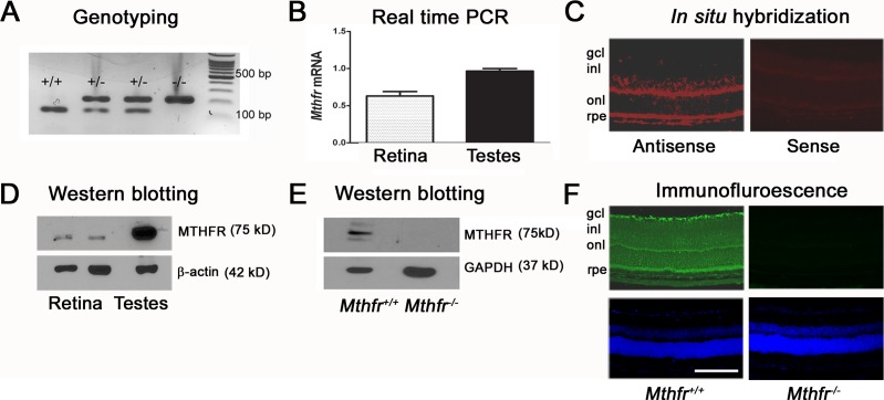
Figure 2.
Assessment of Mthfr gene and protein expression. (A) Representative genotyping data for four offspring; wild type mice expressing only the 145-base pair (bp) product have both copies of the Mthfr gene, heterozygous mice expressing the 216-bp and 145-bp have one copy of the Mthfr gene, and homozygous mice expressing only the 216-bp product do not express the Mthfr gene. (B) Total RNA was isolated from neural retina and testis (positive control) and subjected to qRT-PCR with mouse-specific Mthfr primers. The GAPDH served as the internal control. (C) Retinal Mthfr mRNA localization was investigated by FISH. Retinal cryosections were incubated with Mthfr anti-sense and sense probes. Red fluorescence represents positive staining. (D) Protein was extracted from Mthfr+/+ mouse retina and testis, and subjected to immunoblotting with antibody against Mthfr (Mr = 5 kD). The β-actin (Mr = 42 kD) was the internal loading control. (E) To confirm the specificity of the Mthfr antibody, protein was extracted from Mthfr+/+ and Mthfr−/− retina and subjected to immunoblotting with the Mthfr antibody. The GAPDH (Mr = 37 kD) was the internal loading control. (F) Retinal cryosections from Mthfr+/+ and Mthfr−/− mice were incubated with an antibody against Mthfr followed by incubation with Alexa Fluor 488 (green)–labeled secondary antibody. We used 4′6-dimidino-2-phenylindole (DAPI) to label nuclei (blue fluorescence). Calibration bar: 50 μM. gcl, GCL; inl, INL; onl, ONL; rpe, RPE.

