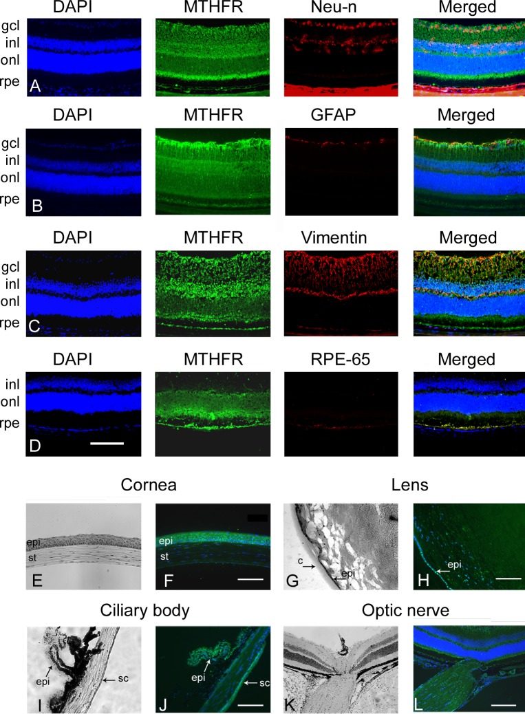
Figure 3.
Immunofluorescent co-localization of Mthfr in retina and in ocular tissues. Retinal cryosections prepared from Mthfr+/+ mice were co-immunostained with antibody against Mthfr (green fluorescence) and antibodies against various cell types (red fluorescence). (A) Neu-N to label retinal neurons of gcl and inl. (B) GFAP to label astrocytes. (C) Vimentin to label Müller cells. (D) RPE-65 to label RPE cells. DAPI was used to label nuclei (blue fluorescence). (E) Eye cryosections prepared from Mthfr+/+ mice were immunostained with antibody against Mthfr (green fluorescence). Companion sections were stained with hematoxylin and eosin. Mthfr was detected in cornea (E, F), lens (G, H), ciliary body (I, J), and optic nerve (K, L). Calibration bar: 50 μM (A–J) and 100 μM (K–L). We used DAPI to label nuclei (blue fluorescence). epi, epithelium; st, stroma; c, capsule; sc, sclera.

