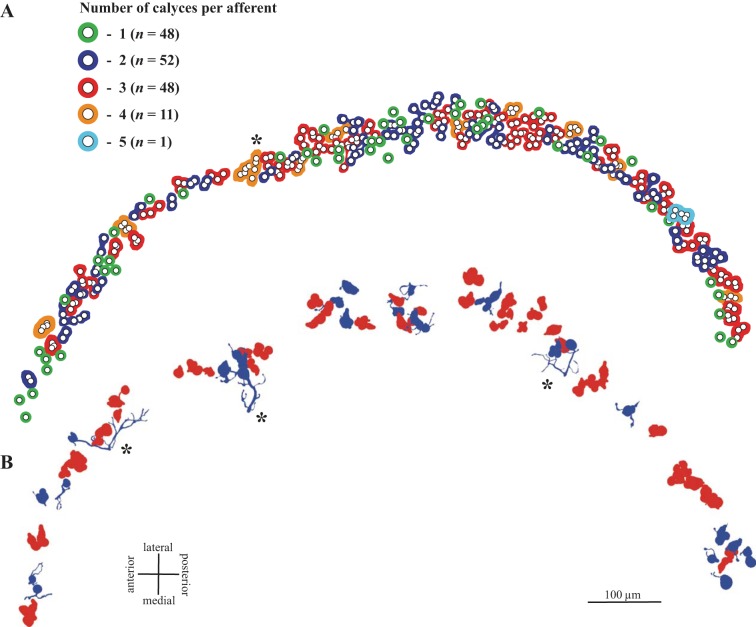
Fig. 3.
CD units in turtle macula. A: calyx complexity. Calyces from all CD units in 1 β-III tubulin-peroxidase-labeled case are indicated by small circles (corresponds to case 1 in Table 2). Colored profiles surrounding 1 or more circles demarcate all calyces arising from a single afferent; colors indicate the number of calyces arising from that afferent (1–5; see key). Note that units supply a restricted region of the striola; they do not extend longitudinally along the striola or, with rare exceptions (*), span the mediolateral width of the striola. B: reconstructed CD units: approximate locations of the 25 D units (blue) and 43 C units (red) reconstructed in this study. Note that most D units, like C units, have restricted collecting areas. Asterisks mark 3 exceptions; these are the only D units with >10 boutons.

