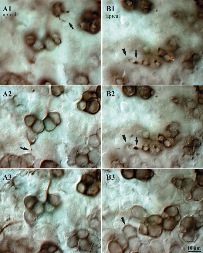
Fig. 6.
Spines on CD units. A and B: DIC micrographs showing examples of spines that arise from BDA-peroxidase-labeled calyces and terminate near the apical surface of the macula. The 3 images in each series (A1–A3, B1–B3) show 3 focal planes from most apical (1) to most basal (3); vertical steps between images are 3–10 μm. A1: 2 spines arise from a labeled calyx and terminate near the apical surface of the epithelium (arrow); under DIC optics they appear to contact the necks of unlabeled hair cells. A2: a third spine arises from the neck of a labeled calyx (arrow); the base of this calyx is visible in A3. B1: a spine with a relatively large terminal swelling (arrow) terminates just above the neck of a calyx (double arrowhead). B2: the same contact seen slightly deeper in the epithelium; the body of the calyx (double arrowhead) is clearer than in B1. B3: base of the same calyx. Such images suggest that spines terminate near the apices of type I hair cells.

