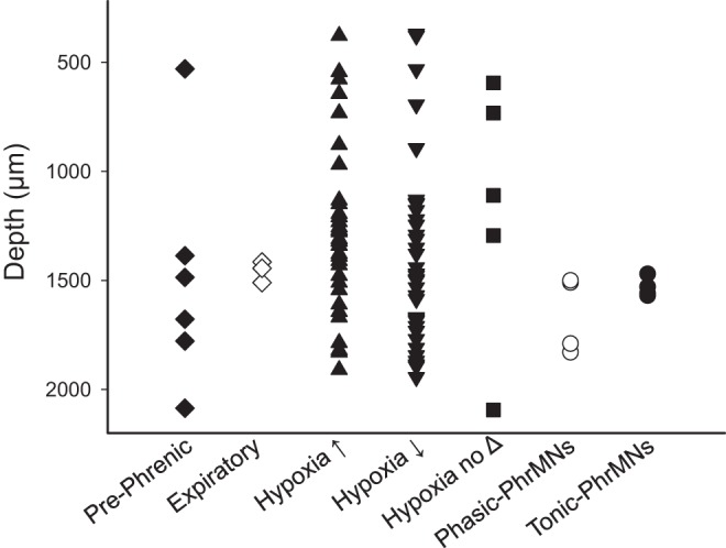
Fig. 5.

The distance (μm) from the dorsal surface of the spinal cord at which each cell was recorded. The microelectrode array was inserted into the spinal cord at 700 μm lateral to the midline at the C4 segment. The classification of cells into groups is discussed in the text as well as Fig. 4 legend. Phasic and tonic phrenic motoneurons (PhrMNs) were recorded at locations consistent with the anatomical location of the phrenic motor pool (i.e., 1,500–1,800 μm below the surface). Most cells classified as “prephrenic” were also recorded in this region, with one exception. Interneurons which responded to hypoxia with increased (hypoxia↑), or decreased (hypoxia↓) burst activity were recorded throughout the entire range of the recording tract.
