
Fig. 6.
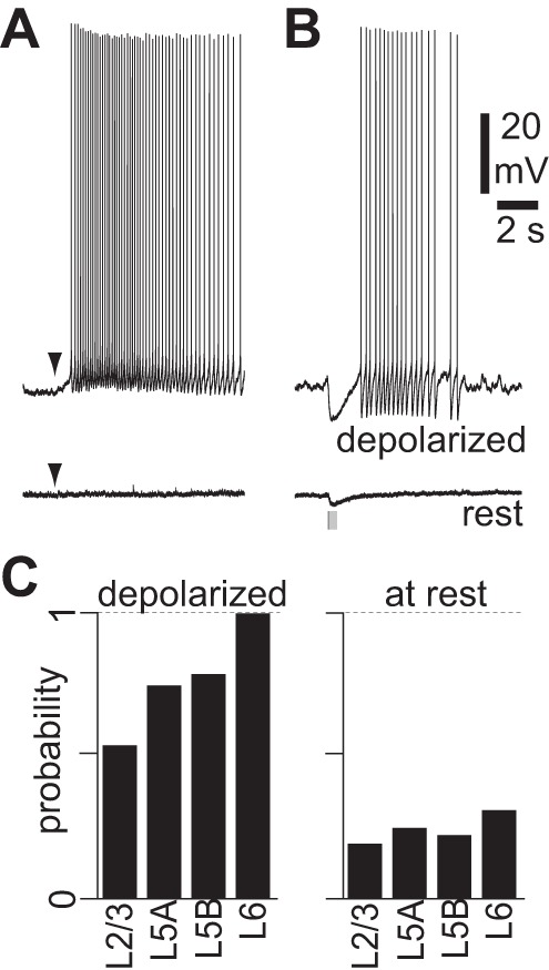
mAChR-mediated hyperpolarization and slow depolarization. A: example of pressure application of ACh (100 μM, 10 ms, arrowheads) at rest and during constant depolarization in a neuron displaying a mAChR-mediated slow depolarization. NBQX, CPP, gabazine, and CGP 52432 were present to prevent network effects. B: example of blue light stimulus at rest and during constant depolarization in a neuron from a virus-injected mouse displaying a mAChR-mediated hyperpolarization and slow depolarization. Stimulus: 10 × 5-ms illumination at 20 Hz. NBQX, CPP, gabazine, and CGP 52432 present throughout. C: incidence of light-evoked mAChR-mediated responses in pyramidal neurons in different cortical layers at rest and during depolarization by somatic current injection. Probability was calculated as the number of neurons in which we observed mAChR responses divided by the total number of recordings. Laminar locations determined by distance from pia (see methods). Depolarized: layer(L)2/3, 8 neurons; L5A, 8 neurons; L5B, 48 neurons; L6, 7 neurons. At rest: L2/3, 22 neurons; L5A, 8 neurons; L5B, 84 neurons; L6, 32 neurons.