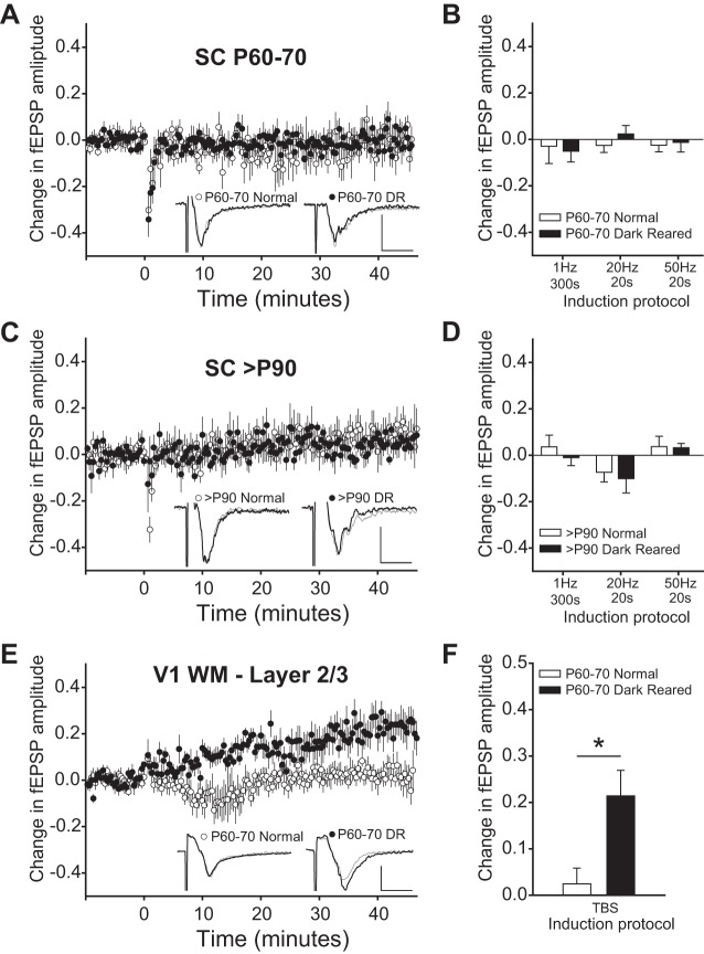
Fig. 2.
Long-term plasticity in SC is not affected by visual experience. A: fEPSPs were evoked at 20-s intervals. After the fEPSP amplitude was stable for >10 min, a 50-Hz, 20-s stimulus was applied. There was no significant change in slices from normal or from dark-reared (DR) animals between postnatal days 60–70 (P60-70). Example traces are averages during a 10-min baseline (thin gray) and 30–40 min after the induction protocol (thick black). Scale bar: 0.2 mV, 5 ms. B: other induction protocols at this age also did not cause long-term plasticity. Data plotted are mean change from 30–40 min after the induction stimulus normalized to a 10-min baseline. C: there was also no significant change after a 50-Hz, 20-s stimulus in slices from normal or DR animals >P90. Example traces and scale bar are as in A. D: there were also no differences between slices from normal or DR animals using 3 other induction protocols. E: in contrast, in primary visual cortex (V1) white matter (WM)-layer 2/3 long-term potentiation (LTP) was induced in slices from DR animals, but not in slices from normal animals. Example traces and scale bar are as in A. F: theta burst stimulation (TBS) caused significantly more LTP in V1 slices from DR than from normally reared animals. *P < 0.05.

