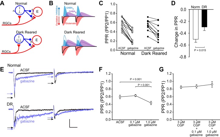
Fig. 5.
Reduced GABAAR function underlies increased PPD in slices from DR animals. A: hypothetical circuit model showing that, when GABAARs are reduced by DR (or blocked), the inhibitory neurons (I) are disinhibited, and GABA release increases, resulting in increased GABABR-mediated inhibition of excitatory neurons (E). Triangles are excitatory, circles are inhibitory. RGCs, retinal ganglion cells. B: model showing how reduced GABAAR function, increased GABA release, and increased GABABR-mediated inhibition could increase PPD in slices from DR animals. Increased GABA release enhances GABABR-mediated inhibition of excitatory neurons (E), which increases the fEPSP duration, and increases inhibition of PP2 (dotted lines are normal condition for comparison). C and D: the GABAAR antagonist gabazine (10 μM) reduced the PPR significantly more in slices from normal animals (50.3 ± 5.08% decrease in PPR) than in slices from DR animals (29.6 ± 6.04% decrease in PPR). E: gabazine caused a large increase in the duration of the fEPSP in slices from both normal and DR animals, which is likely caused by disinhibition of both excitatory and inhibitory neurons and increased duration of evoked responses. Stimulus artifacts are omitted for clarity. Scale bar: 0.2 mV, 20 ms. Arrows indicate peaks of fEPSPs. F: 1 μM gabazine was sufficient to increase PPD (decrease PPR) in slices from normal animals. Thus reducing GABAAR function in slices from normal animals mimicked the increased PPD seen in slices from DR animals (cf., Fig 3). G: blockade of GABABRs prevented the increase in PPD caused by 1 μM gabazine, supporting the model in which reduced GABAAR function increases PPD by increasing inhibitory neuron output and, in turn, increasing GABABR-mediated inhibition.

