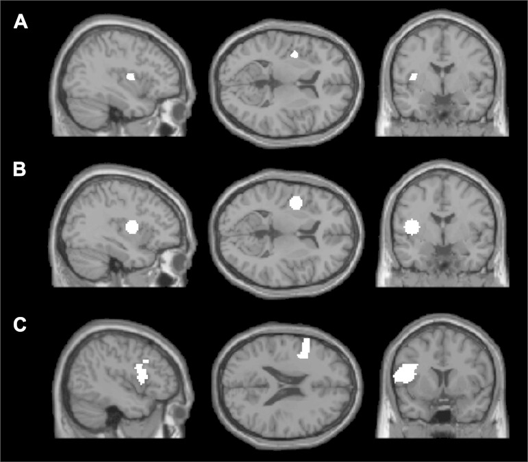
Fig. 1.
The anatomical region of interest (ROI) masks, shown on 3 sample slices on the Montreal Neurological Institute (MNI)152 brain template: A: superior precentral gyrus of the insula (SPGI) mask hand-created by Dronkers and colleagues. B: SPGI mask created by drawing a sphere (radius = 10 mm) around the peak coordinate reported in Dronkers (1996). C: opercular portion of the left inferior frontal gyrus (LIFG). Each mask was intersected with each subject's activation map for the hard articulation > fixation contrast, and the top 10% of voxels in each mask were taken as that subject's functional ROI.

