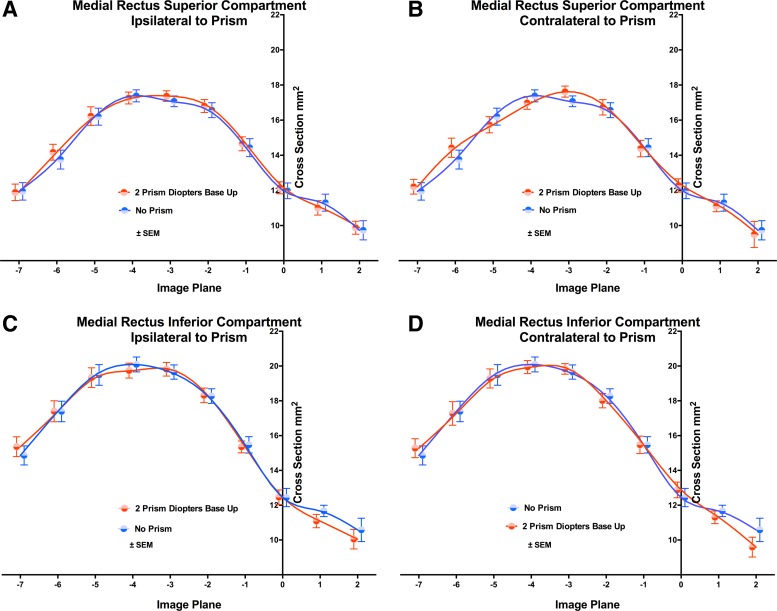
Fig. 5.
Cross-sectional area distributions of the MR muscle in 2-mm-thick image planes numbered as in Fig. 2. A and B: superior compartment. C and D: inferior compartment. A and C: ipsilateral to prism. B and D: contralateral to prism. Viewing through two PD BU prism had no effect on the cross-sectional distribution of either compartment ipsilateral or contralateral to the prism. Symbols and spline fits have been offset slightly on the abscissa to avoid overlap.

