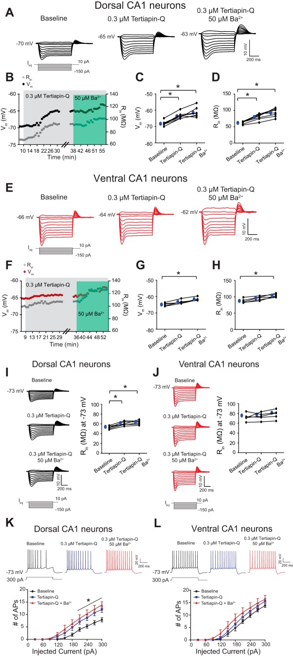
Fig. 3.
Ba2+-Sensitive conductance-mediated changes in Vm and Rin stem from GIRK conductance in dorsal CA1 neurons. A and E: representative voltage responses with step current commands at RMP are shown. B and F: time courses of changes in Vm and Rin during successive 0.3 μM Tertiapin-Q and 50 μM Ba2+ application in dorsal (B) and ventral (F) CA1 pyramidal neurons. C and D: dorsal CA1 neurons showed a significantly depolarized Vm (C) and an increased Rin (D) following bath application of Tertiapin-Q (0.3 μM). Subsequent blockade of Ba2+-sensitive conductance showed no significant changes in Vm (C) and Rin (D) in dorsal CA1 neurons. G and H: ventral CA1 neurons showed no significant changes in Vm and Rin in the presence of Tertiapin-Q (0.3 μM). Further changes in Vm and Rin were not observed following blockade of Ba2+-sensitive conductance in ventral neurons. Blockade of Tertiapin-Q and Ba2+-sensitive conductance produced a significantly depolarized Vm and increased Rin compared with baseline in ventral neurons. I and J, left: representative voltage responses with step current commands at −73 mV. Dorsal CA1 neurons showed a significantly increased Rin (I) but not ventral CA1 neurons (J) at a common Vm (−73 mV) following successive Tertiapin-Q and Ba2+ application. K and L: representative Vm responses with depolarizing current steps (300 pA for 750 ms) at a common Vm (−73 mV). K: dorsal neurons showed significant increases in action-potential (AP) firing after either Tertiapin-Q or Tertiapin-Q + Ba2+ application but not ventral neurons (L). *P < 0.05.

