Fig. 3.
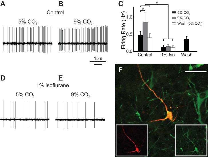
Isoflurane abolished the change in firing rate of a 5-HT neuron in situ in response to acidosis. A: spontaneous firing of a putative 5-HT neuron exposed to control perfusate (pH 7.4) in situ. B: firing frequency increased in response to hypercapnic perfusate (pH 7.2). C: summary of recordings testing 5-HT neuron chemosensitivity in situ (n = 9). 5-HT neurons were chemosensitive under control conditions (P = 0.03). Isoflurane significantly reduced firing frequency (P = 0.02) and caused loss of the response to hypercapnic perfusate. Firing frequency returned to baseline levels after isoflurane was washed out. Error bars represent SE. D: isoflurane (1%) caused a decrease in firing frequency of the neuron in A. E: in isoflurane, the neuron no longer responded to hypercapnic perfusate with an increase in firing frequency. F: juxtacellular labeling confirmed that this was a 5-HT neuron. Red, biotinamide; green, TpOH immunostaining; yellow, colocalization. Scale bar, 50 μm. *P < 0.05.

