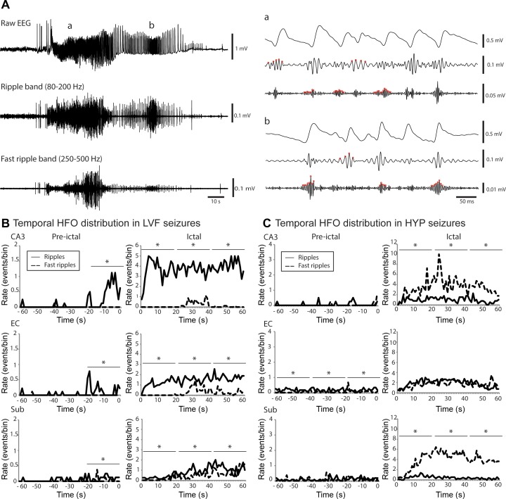
Fig. 2.
High-frequency oscillations (HFOs) during seizures after the injection of 4AP and picrotoxin. A: raw EEG recordings of a HYP seizure recorded from the subiculum in a picrotoxin-treated rat as well as filtered traces in the ripple frequency band (80–200 Hz) and fast ripple frequency band (250–500 Hz). Expanded time scale of the selected part (a and b) of raw EEG recording, ripple frequency band, and the fast ripple frequency band of the seizure shown in A. B: temporal distribution of HFOs in all LVF recorded seizures from 4AP-treated animals in the hippocampal CA3 region, the entorhinal cortex, and the subiculum. Left, temporal evolution of HFOs 60 s before the seizure onset; right, distribution from the onset to 60 s after. Note that all regions show higher rates of ripples compared with fast ripples in the 20 s time period that preceded the onset of the seizure and during the seizure (*P < 0.05). C: temporal distribution of HFOs in all HYP recorded seizures from picrotoxin-treated rats in the CA3, the entorhinal cortex, and the subiculum. Note that the CA3 region and subiculum show higher rates of fast ripples compared with ripples during the 60 s time-period that followed seizure onset (*P < 0.05) as well as that there was no significant difference during the ictal period between the rate of occurrence of ripples and fast ripples in the entorhinal cortex. Note the difference in the scale of pre-ictal and ictal period.

