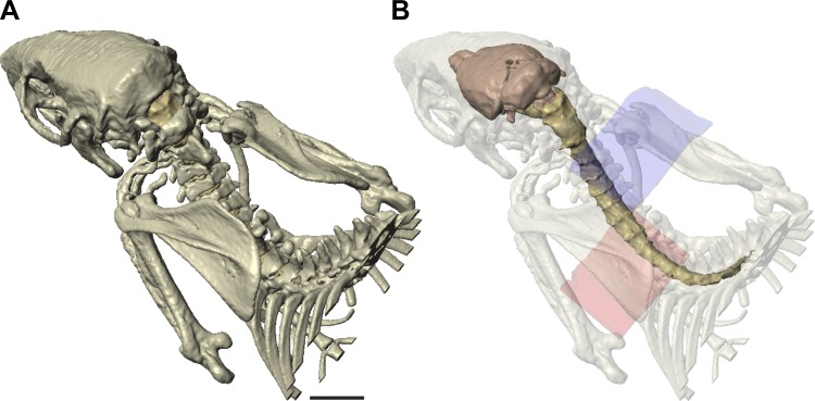
Fig. 1.
High-resolution radiological imaging of the rat and tissue segmentation. A: rendering of bones imaged and segmented for the model. B: rendering of the brain and spinal cord within translucent bone. The cathode (blue, dorsal) and anode (pink, ventral) are shown. Length calibration: 10 mm for A and B.

