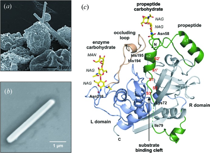
Figure 2.
In vivo grown crystals and three-dimensional structure of the T. brucei cathepsin B–propeptide complex. (a) Scanning electron micrograph of a group of Sf9 insect cells infected with TbCatB virus 80 h after infection showing crystals of overexpressed TbCatB. (b) Scanning electron micrograph of a single TbCatB crystal after isolation. (c) Cartoon plot of the TbCatB–propeptide complex exhibiting the typical papain-like fold of cathepsin B-like proteases. Gray, R domain; blue, L domain; beige, occluding loop. The native propeptide (green) blocks the active site. Two N-linked carbohydrate structures (yellow) consist of N-acetylglucosamine (NAG) and mannose (MAN) residues (yellow, carbon atoms; blue, nitrogen atoms; red, oxygen atoms). [From Redecke et al. (2013 ▶), Science, 339, 227–230. Reprinted with permission from AAAS.]

