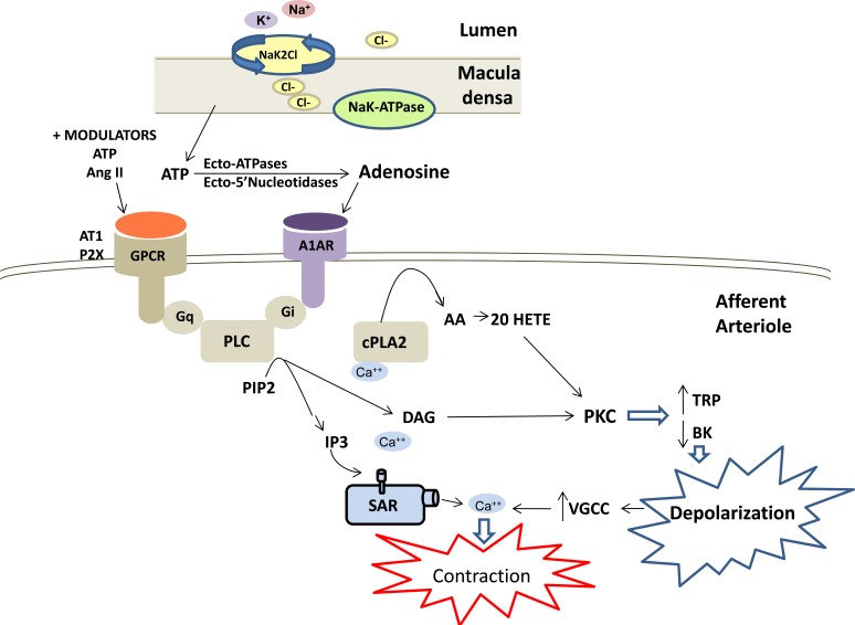
Fig.(3).
Mechanisms involved in tubuloglomerular feedback control of renal vascular tone. Tubuloglomerular feedback is initiated when the filtered load of sodium chloride is increased to the macula densa resulting in increases of; sodium reabsorption via the sodium potassium 2 chloride exchanger (NaK2Cl), intracellular Na+, sodium potassium ATPase (NaK-ATPase) activity and intracellular Ca2+. These events trigger the release of ATP from the macula densa cells. ATP is converted to adenosine extracellularly and then acts on adenosine 1A receptors (A1AR) in the adjacent afferent arteriole to promote vasoconstriction. A1AR activates phospholipase C (PLC) via a GTP binding protein (Gi), the release of inositol trisphosphate (IP3) and diacylglycerol (DAG). The subsequent activation of cytosolic phospholipase A2, formation of arachidonic acid (AA) and 20-HETE, as well as activation of protein kinase C (PKC), alters transient receptor potential channel (TRPC) and large conductance calcium activated potassium channel (BK) activities resulting in depolarization and activation of voltage gated calcium channels (VGCC) and Ca2+ influx. The increase in intracellular Ca2+ initiates contraction of the afferent arteriole, reduction in glomerular capillary pressure and filtered sodium chloride load. Activation of the angiotensin II receptor type I (AT1R) stimulates a similar transduction pathway to that shown in (Fig. 2) for the myogenic response. In addition, ATP and angiotensin II have been shown to be positive modulators of tubuloglomerular feedback through activation of G-protein coupled receptors, specifically purinergic receptor 2X (P2X) and angiotensin 1 receptor (AT1) via Gq to modulate PLC activity.

