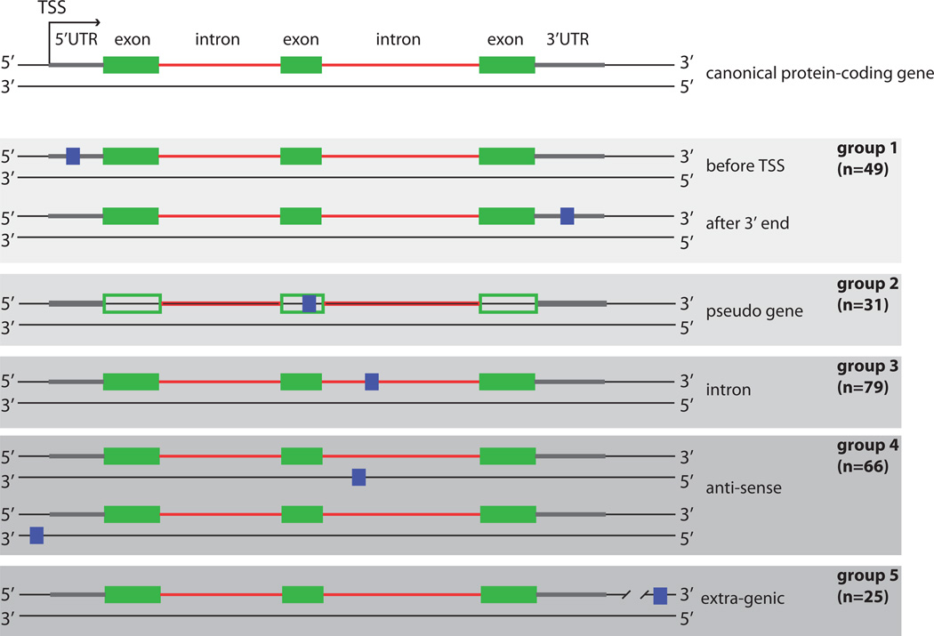
Figure 3.
Classification of novel peptides based on their genomic location. The top panel depicts the two DNA strands of a canonical protein-coding gene with exons (green boxes), introns (red lines), and the transcribed sequences before the first exon and after the last exon (gray lines, here referred to as 5’UTR and 3’UTR). The 250 novel peptides were divided into 5 groups based on their location relative to known genes. The peptide location is denoted here by blue boxes. The number of peptides in each group is indicated. In the last group (extra-genic) the line is cut to indicate that there is a large distance (>10 kb) from the protein-coding gene. TSS = transcription start site.

