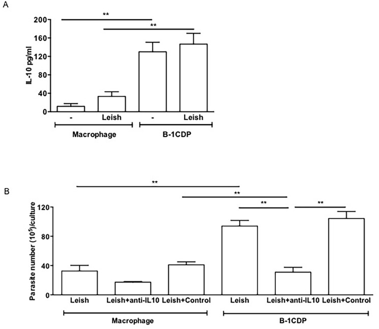
Fig 2. IL-10 is determinant for the increased parasite load in B-1CDP.
B-1CDP cells and peritoneal macrophages were cultured in the presence or absence of L. major (MOI 10:1). After 24 hours, the supernatant was collected and IL-10 (A) was measured by ELISA. All cultures were performed in triplicate and bars show the mean ± SD. Representative result of three similar experiments **p <0.05. B-1CDP cells and peritoneal macrophages were treated or not with doses of monoclonal neutralizing anti-IL-10 or control isotype. Once were infected with L. major, after 24 hours, the cell cultures were washed with DMEM and incubated 3 days and then passed to Schneider medium. After 5 days in medium Schneider, promastigotes were counted in the supernatant (B). Statistical analysis were performed by Student’ t test from representative results of three similar experiments and bars show the mean +SD. **p ≤ 0.05.

