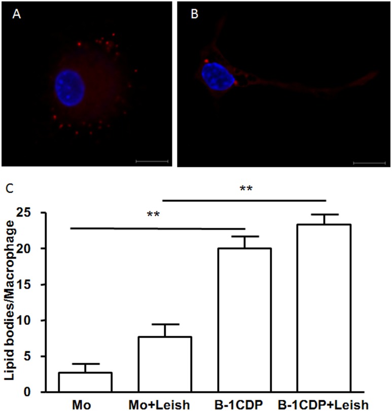
Fig 3. B-1CDP cells naturally express increased numbers of lipid bodies as compared to peritoneal macrophages.
B-1CDP cells (A) and peritoneal macrophages (B) were incubated with glass coverslips, some cultures were infected with L. major (3C). Stained with Nile red (Sigma), the slides were washed and stained with DAPI (Sigma). The morphology of fixed cells was observed, and Nile red LBs were counted by light microscopy with a 100× objective lens in 50 consecutively scanned leukocytes. Statistical analysis were performed by Student’ t test from representative results of three similar experiments and bars show the mean +SD. **p ≤ 0.05. Bar, 10 μm. Representative of two experiments with identical results.

