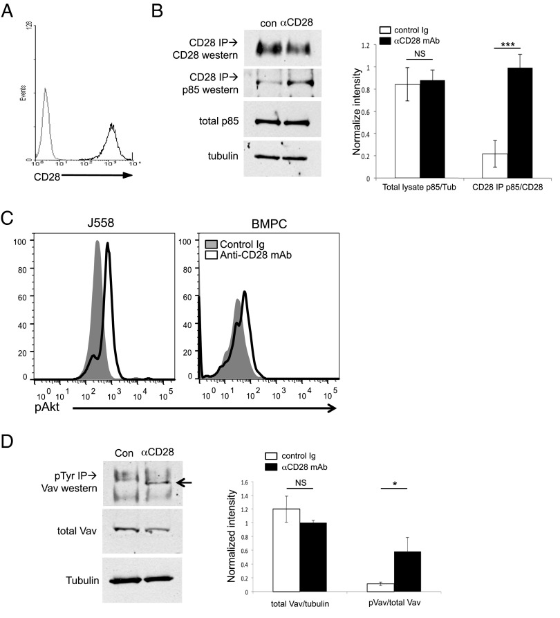
FIGURE 1.
CD28 activation induces PI3K and Vav signaling in PC. J558 murine plasmacytoma cells were activated with control hamster Ig (con) or anti-CD28 mAb (αCD28, PV1.1) for 15 min and then analyzed by coimmunoprecipitation, followed by Western blot. (A) CD28 expression by J558, by flow cytometry. (B) PI3K. Left, Protein lysates from J558 activated as indicated were immunoprecipitated with αCD28 mAb, and the immune complexes were analyzed by Western blot for coimmunoprecipitation of the p85 subunit of PI3K. Total p85 and tubulin (as a loading control) were determined by Western blot from total cell lysates. Data representative of three independent experiments. Right, Relative densitometry combined from three experiments. (C) Downstream AKT activation. J558 cells (left) or purified BM PC (right) were treated with control Ig (filled histograms) or αCD28 mAb (open histograms) for 1 h in serum-free media, fixed, stained for phospho-Akt, and analyzed by flow cytometry. Data representative of two independent experiments. (D) Vav. Protein lysates from J558 activated as indicated were immunoprecipitated for phosphotyrosine, and the immunocomplexes were analyzed by Western blot for the presence of Vav1 (arrow). Data are representative of three independent experiments. Right, Relative densitometry combined from three experiments. The same results were seen when the Western blot was probed for phospho-Vav1 (data not shown). *p < 0.05, ***p < 0.001.

