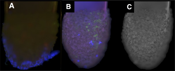
Figure 1.

Tano scraper tips show ILM fragments and cells. After they were used for treatment of patient retinas during macular hole surgery, the Tano scratchers were visualized with immunohistochemistry. (A) Numerous cells (blue) can be seen on sheets of tissue attached to the instrument tips. (B) Laminin A–positive tissue fragments (green) indicate ILM and cells (blue) on the Tano tips. (C) Control confocal fluorescence image of an unstained tip.
