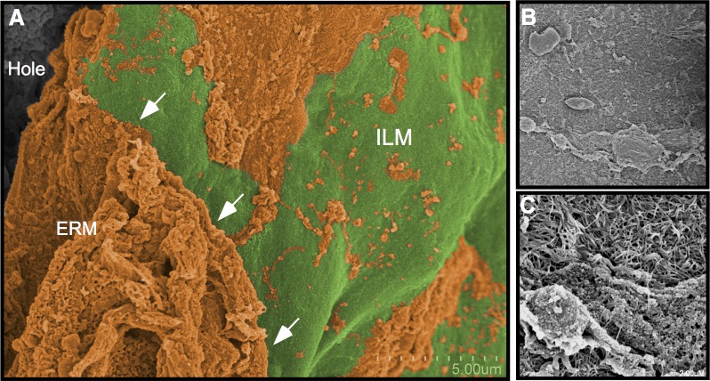
Figure 4.
Area 2: Macula with light Tano application. (A) After a light application of the Tano, an edge (arrows) of ILM (green) becomes apparent alongside a region where untreated ERM (orange) remains. (B, C) Higher magnification over the scraped area shows that some cell and tissue elements of the epiretinal membrane remain on the surface of the ILM.

