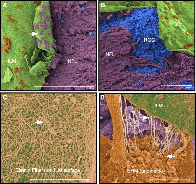
Figure 6.
Area 4: Macula with heavy Tano application. (A, B) Heavy application of the Tano scraper caused a tear in the ILM (green) and subsequent exposure of the nerve fiber layer (purple) and cellular elements (blue). The underside of the ILM (arrow) shows fragments of the retinal nerve fiber layer (purple) remain attached to the ILM. (C) Fibers consistent with elastin (orange) were found between the ERM and ILM. (D) These fibers remain attached to the ILM and underside of the ERM.

