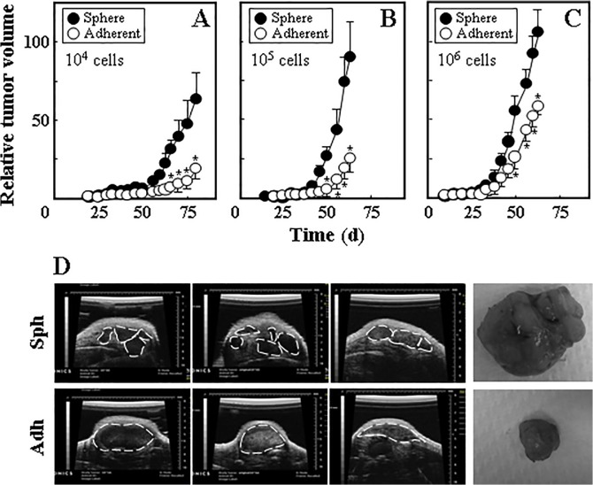
Fig 3. Ist-Mes-2 sphere cells form tumours more efficiently than adherent cells.
Ist-Mes-2 adherent and sphere cells were injected s.c. into Balb-c/nude mice at 104 (A), 105 (B) and 106 cells per mouse (C). Tumour volume was evaluated using USI and is expressed relative to the initially detected tumour (~20 mm3). (D) Three different representative images taken by ultrasound from sphere- and adherent cell-derived tumours are shown for tumours grown from 106 cells per mouse. The broken line indicates contours of the USI-visualised tumours. On the right hand side, a photograph of a representative tumour grown from 106 sphere (upper image) and adherent cells (lower image) is shown. Data in panels A-C are derived from experiments comprising 4–5 mice per group and are mean values ± S.E.M. The symbol ‘*’ indicates significantly different values with p<0.05.

