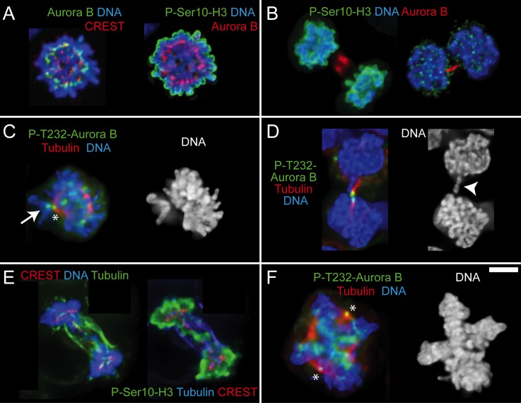
Fig 2. Aurora B activity is normal in cHL small and large cells.
A-F. Immunofluorescence images of small mononucleated cells (A-E) and large cell undergoing mitosis (F). A. Cell in prometaphase with Aurora B kinase properly localized at centromeres and phospho-Ser10-histone H3 on chromatin. B. One cell in telophase (left) and one cell in the last stages of cytokinesis (right). Aurora B localizes properly in the central spindle or in the thin intercellular bridge that connects the two sister cells, respectively. As expected, signal for phospho-Ser10-histone H3 is only observed in the cell on the left where nuclear envelope has not reformed yet. C. Cell in prometaphase with most of the chromosomes aligned at the metaphase plate and one mono-oriented chromosome on the spindle pole with the unattached kinetochore showing prominent accumulation of auto-phosphorylated Aurora B (white arrow). Asterisk indicates a spindle pole that is unspecifically labeled by the P-T232-Aurora B antibody. D. Cell in the last stages of cytokinesis with chromatin present in the intercellular bridge (white arrowhead). Auto-phosphorylated Aurora B accumulates on the intercellular bridge revealing that Aurora B has been properly activated at a time when abscission is taking place. E. Cell in telophase with chromatin bridges staining positive for phospho-Ser10-histone H3. F. Large cell with a double metaphase plate showing auto-phosphorylated Aurora B and therefore active kinase on centromeres. Asterisks are the spindle poles. Size bar is 5 μm.

