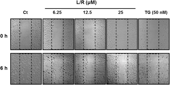
Fig 6. Effect of HIV PIs on cell migration.
HNSCC SQ20B cells were plated on 12-well plates until confluent. Cells were scratched to simulate a wound and images were recorded as “0h”. Cells were treated with different amounts of HIV PIs (L/R, 0–25 μM) for 6 h. The images of wound areas were recoded as described in Methods. Representative images are shown.

