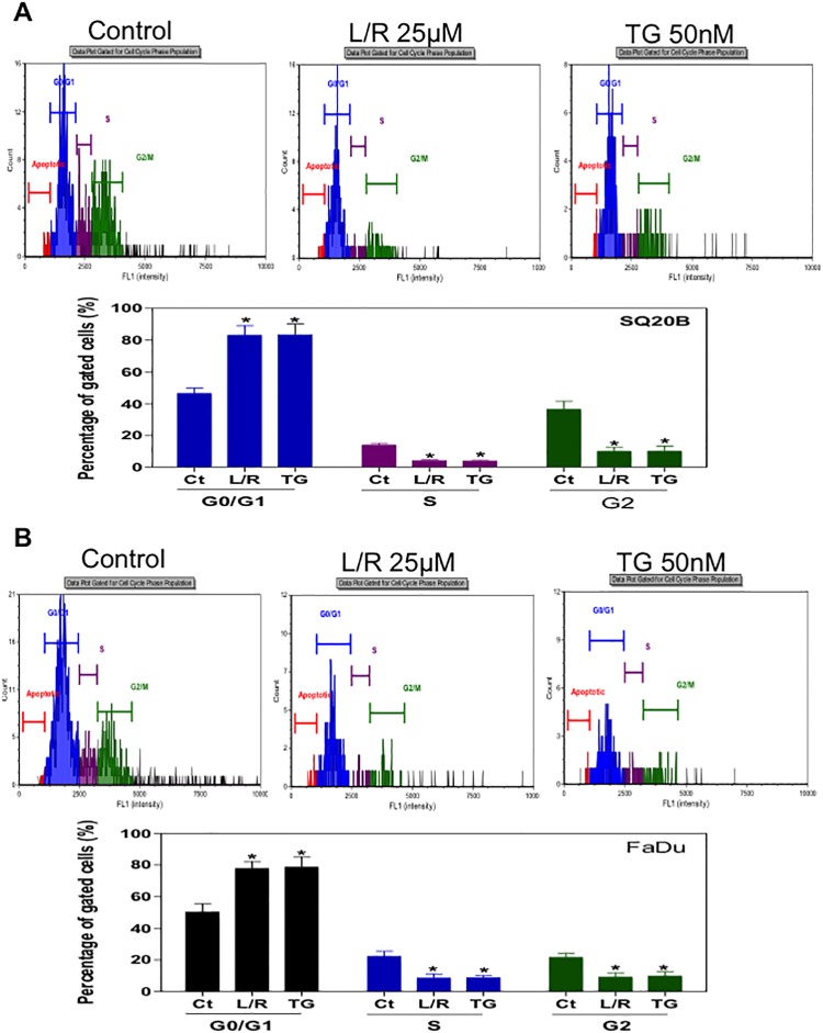
Fig 7. Effect of HIV PIs on cell cycle distribution of HNSCC cells.
Human HNSCC cells (A) SQ20B and (B) FaDu were cultured in complete medium and treated with L/R (25μ M) or TG (50 nM) for 24h. At the end of treatment, cells were stained with the Cellometer PI Cell Cycle kit and analyzed by the Cellometer Vision CBA system. Cell population percentages of G0/G1, S and G2/M phases are shown. Values of each group are mean ± S.E. of three independent experiments. *P<0.05, Statistical significance relative to control group.

