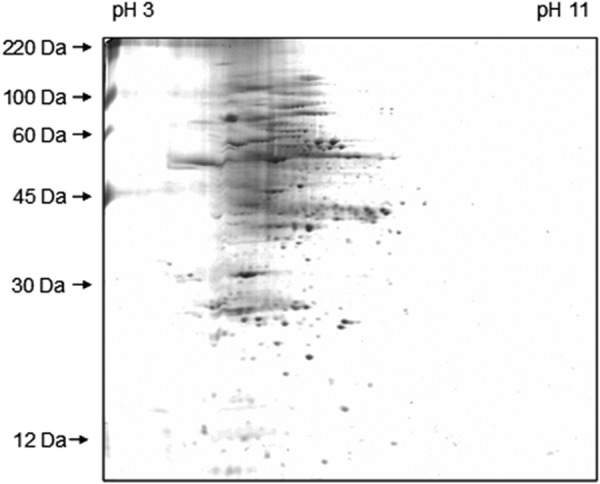
FIGURE 1.

Two-dimensional SDS-PAGE gel of Z. mays L leaf. Separation was achieved by isoelectric focusing (pH 3–11) in the horizontal dimension and by SDS-PAGE in the vertical dimension. The spots were visualized by staining with Coomassie Blue R250.
