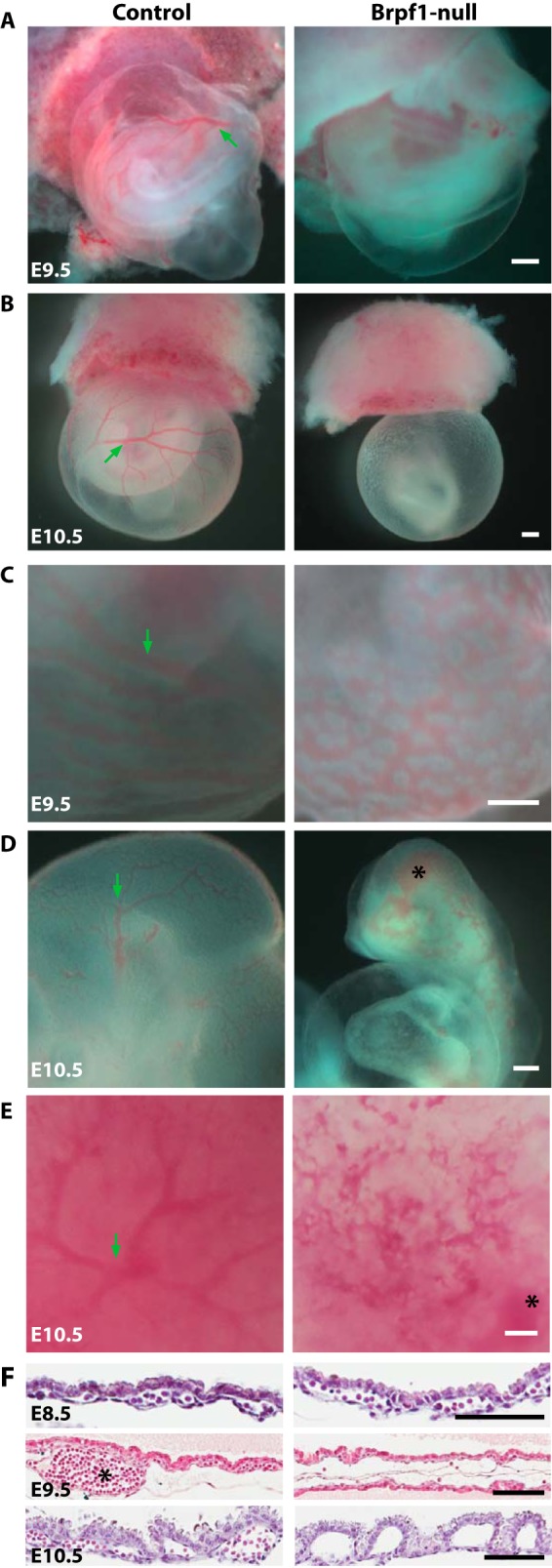
FIGURE 1.

Aberrant blood vessels in Brpf1-deficient conceptuses. A and B, gross morphology of control and knockout conceptuses at E9.5 and E10.5. Note that the mutant yolk sacs were pale, without any visible vasculature. At E8.75, mutant conceptuses appeared normal (data not shown). C, in a less severe mutant yolk sac, a nonhierarchial vasculature was observed at E9.5, compared with the large vessels (indicated by a green arrow) and small branches of the control yolk sac. D and E, arrested vasculogenesis also occurred in the embryo proper (D) and chorionic plate (E) at E10.5. Note the hemorrhage in the mutant cephalic region and chorionic plate, marked with black asterisks in D and E, respectively. F, H&E staining of control and mutant yolk sac sections at E8.5 (top), E9.5 (middle), and E10.5 (bottom). At E8.5, nucleated red blood cells were found in both control and mutant yolk sacs. At E9.5, in those severely affected mutant yolk sacs, only a few erythrocytes could be found in serial sections, whereas in the control yolk sacs, large vessels (marked with a asterisk) were frequently seen. At E10.5, no erythrocytes existed in the mutant yolk sacs. Scale bars, 400 μm (A–E) and 100 μm (F).
