FIGURE 5.
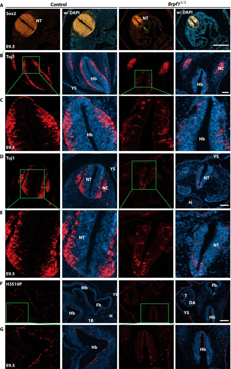
Defective cell proliferation and migration in the mutant neural tube. A, E9.5 embryo sections were immunostained to detect Sox2+ neural precursors. Different from the wild type, the mutant neural tube exhibited uneven distribution of Sox2+ cells. The green arrow marks the loss of Sox2 expression in the ventral part of the neural tube. B and E, anti-Tuj1 immunostaining was performed on transverse sections of control and mutant embryos at E9.5. Boxed regions in B and D are shown in magnified views in C and E, respectively. In the mutant neural tube there were fewer Tuj1+ post mitotic neurons in the cephalic (B and C) and trunk (D and E) regions. In addition, these neurons failed to arrive in the outer epithelium of the neural tube. F, the control and mutant embryos at E9.5 were immunostained to detect Ser-10 phosphorylation of histone H3 (H3S10P). Compared with the control, the mutant embryo contained fewer H3S10P+ nuclei. G, higher magnification images of the hindbrain regions boxed in F. 1B, first branchial arch; DA, dorsal aorta; Fb, forebrain; H, heart; Hb, hindbrain; Mb, midbrain; NC, neural crest; NT, neural tube in the trunk; T, tail; YS, yolk sac. Scale bars, 200 μm in A and F; 100 μm for B and D.

