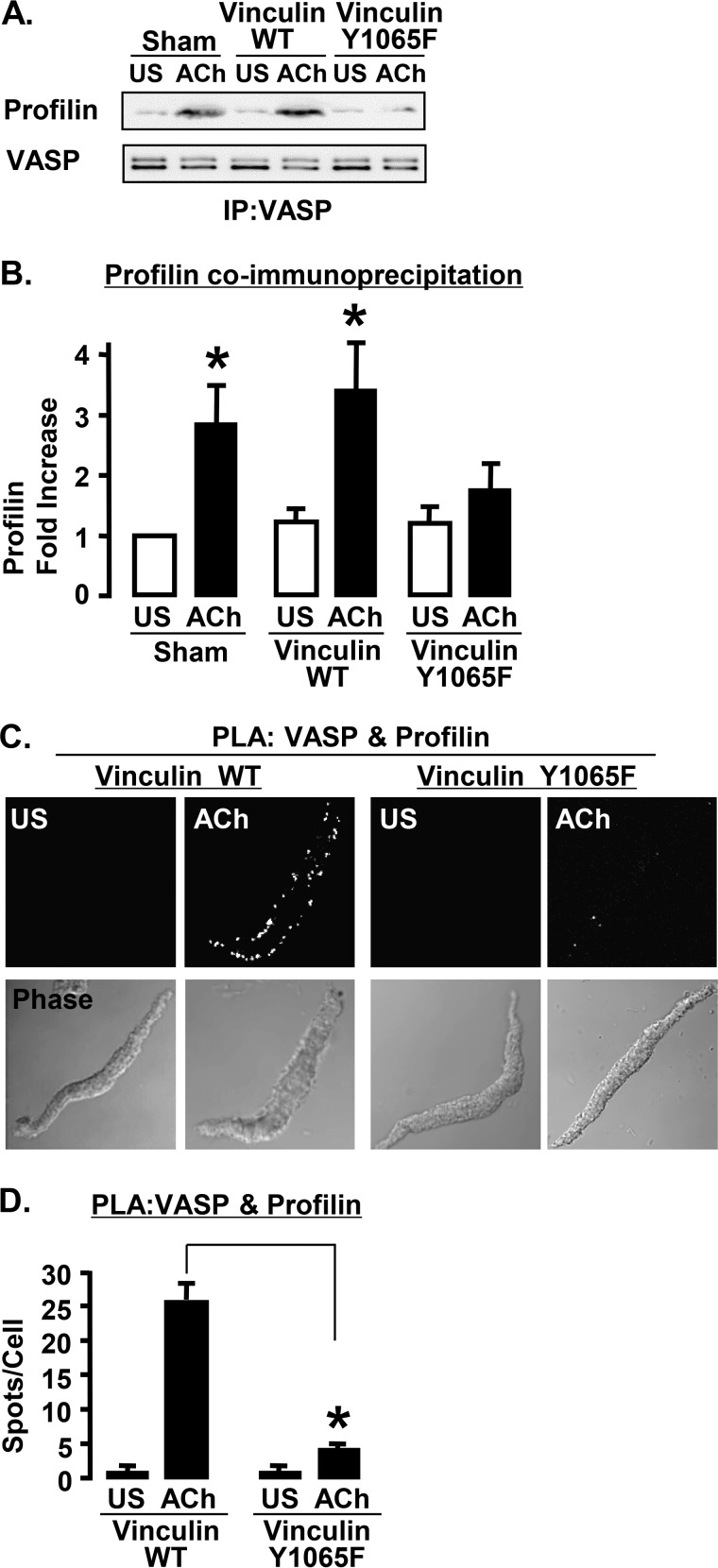
FIGURE 8.
Expression of vinculin Y1065F inhibits the VASP-profilin interaction. A, immunoblots from VASP immunoprecipitates (IP) from tissues expressing vinculin WT, vinculin Y1065F, or sham-treated with or without ACh stimulation. B, the amount of profilin that co-precipitated with VASP during ACh stimulation increases significantly in sham-treated tissues and tissues expressing vinculin WT but not in tissues expressing vinculin Y1065F (n = 5). *, significantly different from US. C, PLA in cells expressing vinculin WT or vinculin Y1065F to visualize the formation of complexes between profilin and VASP. Each fluorescent spot indicates a complex between profilin and VASP. D, *, the total number of PLA spots was significantly higher in ACh-stimulated cells expressing vinculin WT (n = 21) than in cells expressing vinculin Y1065F (n = 21).

