FIGURE 2.
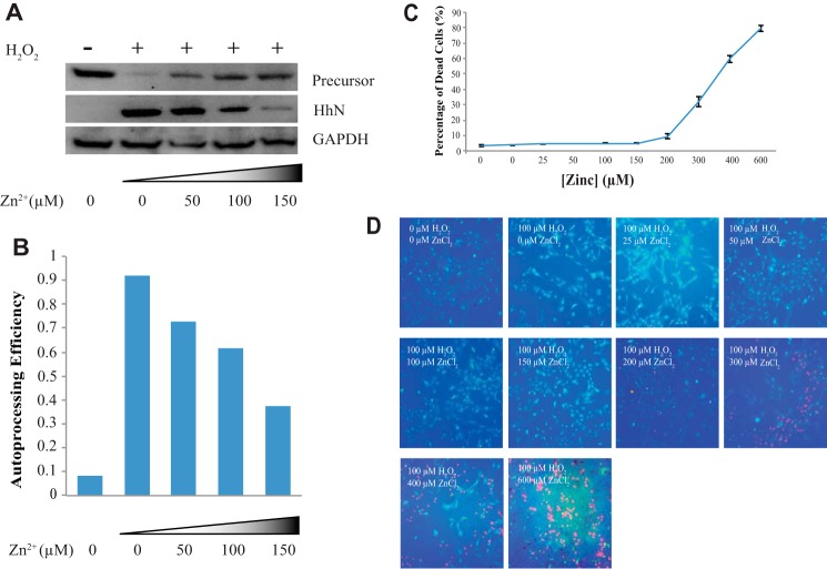
Zinc inhibits Hh autoprocessing in primary astrocyte cell culture. A, zinc inhibits Hh autoprocessing in astrocytes. Immunoblotting for GAPDH was used as a loading control. B, quantitative analysis was performed with ImageJ. The amounts of proteins were normalized to those of GAPDH. C, dose-response curve for percentage of dead rat primary astrocyte cells by varying concentrations of environmental zinc. Cultures were exposed to 0–600 μm zinc and 100 μm H2O2 for 40 h. The cell viability was assessed using the Live/Dead cell imaging kit, and the percentage of dead cells was determined by the Spot Detector BioApplication. Results are expressed as the mean derivations ± S.D. for two cultures. D, a representative figure for each condition. In each figure green fluorescence represents live cells and red fluorescence represents dead cells.

