FIGURE 4.
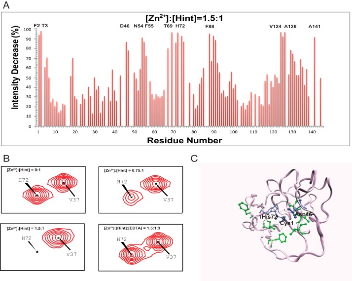
Structural basis of zinc-Hint binding. A, 1H,15N-HSQC signal intensity changes of Zn2+ binding to Hint at 25 °C. The residues with the biggest changes are labeled. B, chemical shift perturbation analysis for His-72 backbone in 15N-labeled Hint upon zinc binding. With increasing amount of zinc, the amide peak intensities of His-72 decreased during the titration. Adding 2 mol eq of EDTA resulted in the complete reappearance of the missing signals. C, structural model of Hint binding site of Zn2+ mapped onto the x-ray structure (PDB ID 1AT0) based on the NMR signal intensity change. Blue residues are the direct coordination sites, which have the biggest signal decrease, whereas green residues, with less signal reduction, likely play a secondary role in zinc binding.

