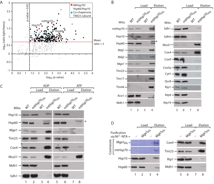
FIGURE 1.
MtHsp70 interacts with the mitochondrial chaperonin system. A, SILAC-based quantitative mass spectrometric analysis of mtHsp70His affinity purifications. The mean log10 light/heavy ratio and −log10 p values of three independent experiments were determined and plotted against each other. The thresholds for the light/heavy ratio >5 and p > 0.05 are indicated. Red dots, mitochondrial chaperones; blue dots, cochaperones of mtHsp70 and TIM23 subunits; black dots, other proteins; gray dots, unspecific proteins (see supplemental Table S1). B, WT and mtHsp70His mitochondria (Mito) were lysed with digitonin and subjected to affinity purification via Ni2+-NTA-agarose. Bound proteins were eluted with imidazole and analyzed by SDS-PAGE, Western blotting, and immunodetection with the indicated antisera. Load, 0.5%; elution, 100%. Aco1, aconitase 1; Mdh1, mitochondrial malate dehydrogenase 1; Sdh, succinate dehydrogenase; Cox, cytochrome c oxidase; Atp, subunits of the F1FO-ATP synthase. The asterisk marks cross-reactivity of anti-Hsp60 antibody with mtHsp70. C, WT and mtHsp70His mitochondria were lysed in digitonin in the presence of ADP or ATP and subjected to affinity purification via Ni2+-NTA-agarose. Bound proteins were eluted with imidazole and analyzed by SDS-PAGE, Western blotting, and immunodetection with the indicated antisera. Load, 0.5%; elution, 100%. The asterisk marks cross-reactivity of anti-Hsp60 antibody with mtHsp70. D, WT mitochondria were lysed in digitonin and incubated with empty or Mge1His-coated Ni2+-NTA-agarose. Bound complexes were eluted with imidazole, and samples were analyzed by SDS-PAGE, Western blotting, and immunodetection with the indicated antisera. Load, 0.5%; elution, 100%.

