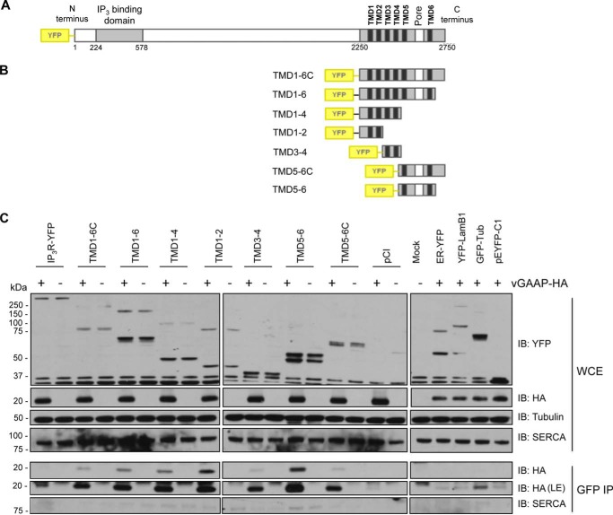
FIGURE 1.
vGAAP is co-immunoprecipitated with all pairs of TMDs from IP3R1. Shown is co-immunoprecipitation (IP) between vGAAP from VACV Evans and full-length and truncated versions of YFP-tagged type 1 IP3R. Shown are schematic representations of the full-length (A) and truncated forms of YFP-IP3R (B) used to map the interaction. C, COS-7 cells were transfected with plasmids encoding the YFP-IP3R proteins, and 18 h later, cells were infected with either v-ΔGAAP (−) or revertant vGAAP-HA (+) VACV and collected after 16 h. Following co-immunoprecipitation with anti-GFP, the immunoprecipitates and the whole cell extracts (WCE) were resolved by SDS-PAGE and immunoblotted (IB) with anti-YFP, anti-HA, anti-SERCA (as a control for contamination with ER and Golgi membrane proteins), and anti-tubulin (loading control) antibodies. Four additional vectors expressing YFP- or GFP-tagged proteins that localize to different organelles were used as negative controls: ER-YFP (ER), YFP-LamB1 (nucleus), GFP-Tub (cytoplasm), and pEYFP-C1 (free YFP). The results shown are typical of three independent experiments. LE, longer exposure.

