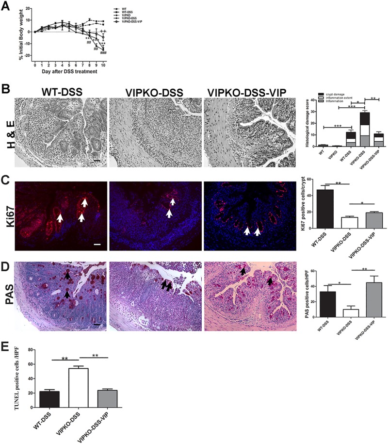
Fig 6. VIP protects mice against DSS-induced colitis.
Exogenous VIP was administered to DSS-treated VIPKO mice daily for 10 days. Change in body weight over the 10-day study period,in naive WT and VIPKO mice, DSS exposed WT (WT-DSS), VIPKO (VIPKO-DSS) and DSS plus VIP treated VIPKO (VIPKO-DSS-VIP) mice (A). Representative H&E stained colon sections and histological damage score at day 10 in DSS exposed WT (WT-DSS), VIPKO (VIPKO-DSS) and DSS plus VIP treated VIPKO (VIPKO-DSS-VIP) mice (B), IEC proliferation determined by Ki67 immunostaining (C), selective labeling of neutral mucins with PAS and quantification of PAS+ve cells as total number of PAS+ve cells/HPF (D) and cell death determined by TUNEL staining (E); n = 6–7 animals/ group, results are represented as means ± SEM, *P<0.05, **P< 0.01, ***P<0.001. Scale bar = 50 μm. Arrows highlight Ki67 +ve cells in (C) and PAS+ve cells in (D).

