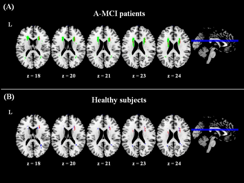
Fig 3. Results of voxel lesion-symptom mapping analyses.
A significant association was found between the anatomical localization of white matter lesion in the anterior thalamic radiation (areas in red) and the presence/severity of apathy in between amnestic mild cognitive impairment (a-MCI) patients (panel A). Voxels in green show the lesion distribution in patients used for the voxel lesion-symptom mapping (VSLM) analysis, i.e., only including areas where at least 4 subjects presented a lesion. The lesion distribution in healthy controls (panel B, in blue), does not overlap with the area found to be significant for apathy in patients. Note that the lesion distribution is thresholded to show only voxels where at least 4 participants had a lesion, in order to match those included in the VLSM analysis.

