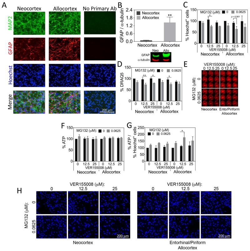
Figure 5. Neo- and allocortical astrocytes do not differ in their reliance on Hsp70/Hsc70 defenses.
(A) Neo- and allocortical (entorhinal) neuronal cultures were stained for MAP2, GFAP, and Hoechst. Some GFAP+ astrocytes were visible in both cultures. Omission of primary antibodies led to loss of fluorescent signal. (B) Western blots for GFAP verify the presence of this protein in cultures from both brain regions and reveal that GFAP protein levels are higher in allocortex cultures. Shown are the mean ± SEM of 6 independent experiments. ** p ≤ 0.01 vs neocortex, two-tailed Student’s t-test. (C–H) Astrocyte cultures harvested from neo- and allocortex were treated with MG132 and the Hsp70/Hsc70 inhibitor VER155008 for 48h and the number of Hoechst+ cells, DRAQ5 nuclear staining, and ATP levels were assessed. ATP levels were also expressed as a function of Hoechst+ cells in panel G. Representative DRAQ5 and Hoechst images from the data in panels C and D are shown (E, H). Unlike in neurons, there was no robust exacerbation of MG132 toxicity in neo- or allocortical astrocytes after loss of Hsp70/Hsc70 activity, even at higher concentrations of VER155008. Shown are the mean ± SEM of 3 independent experiments. + p ≤ 0.05, ++ p ≤ 0.01 vs 0 μM MG132, Bonferroni post hoc correction following three-way ANOVA.

